


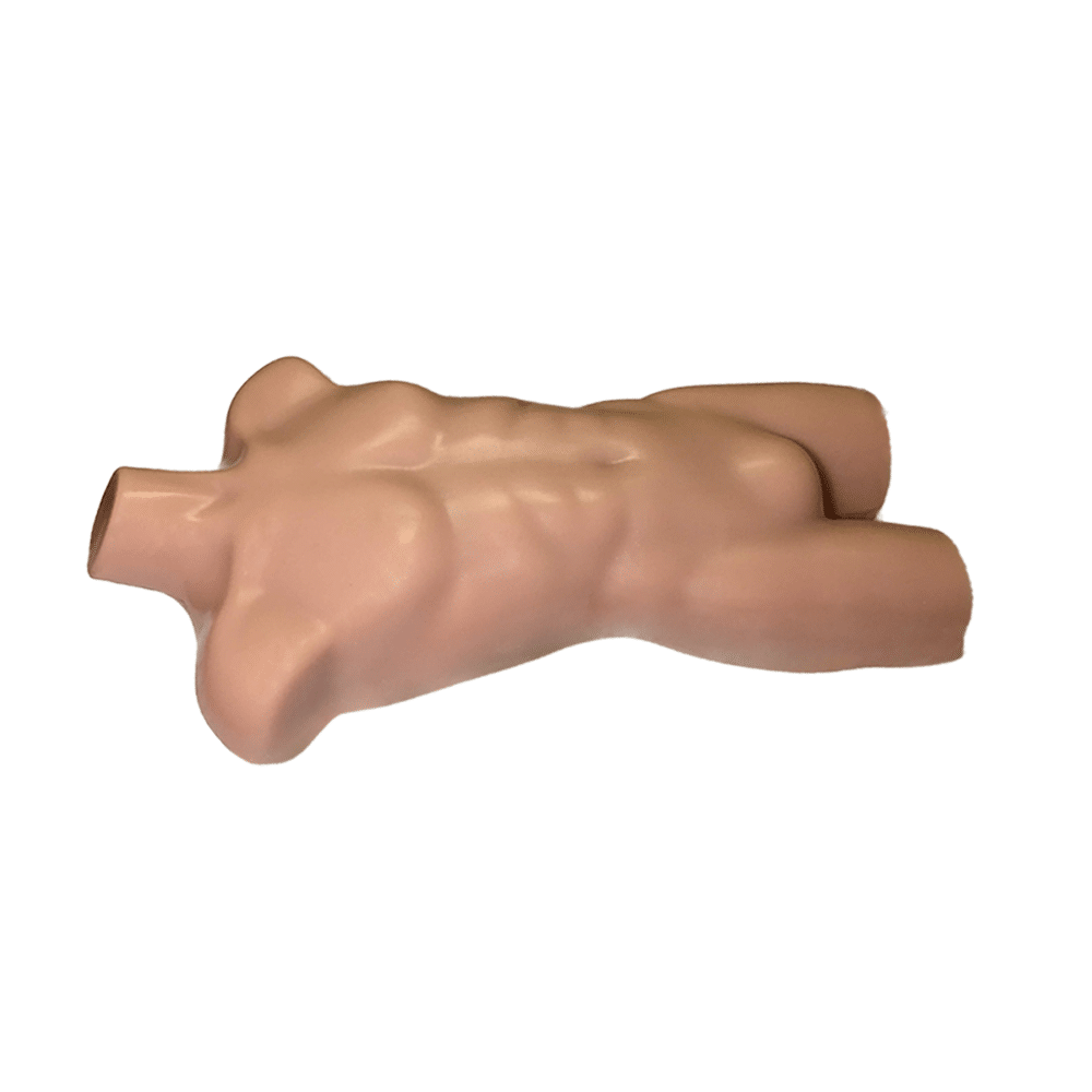
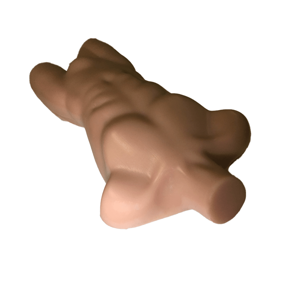
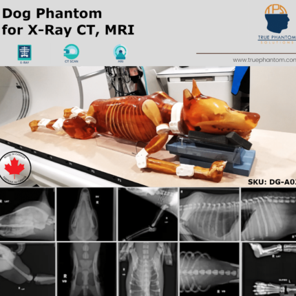
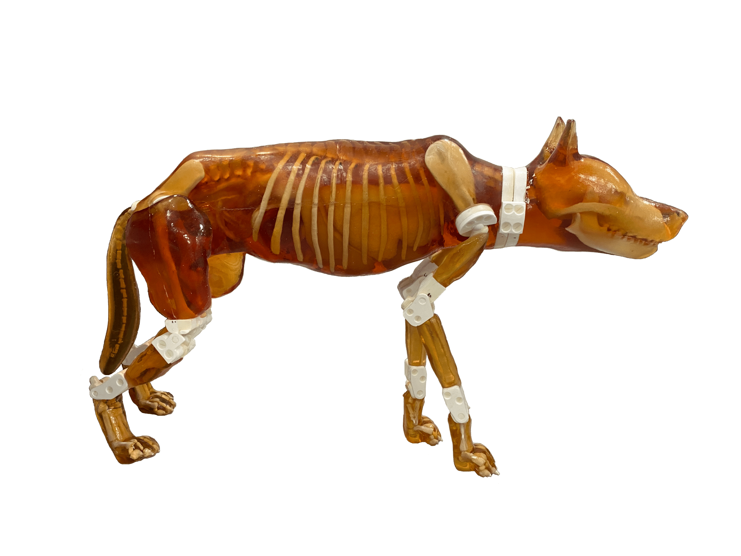
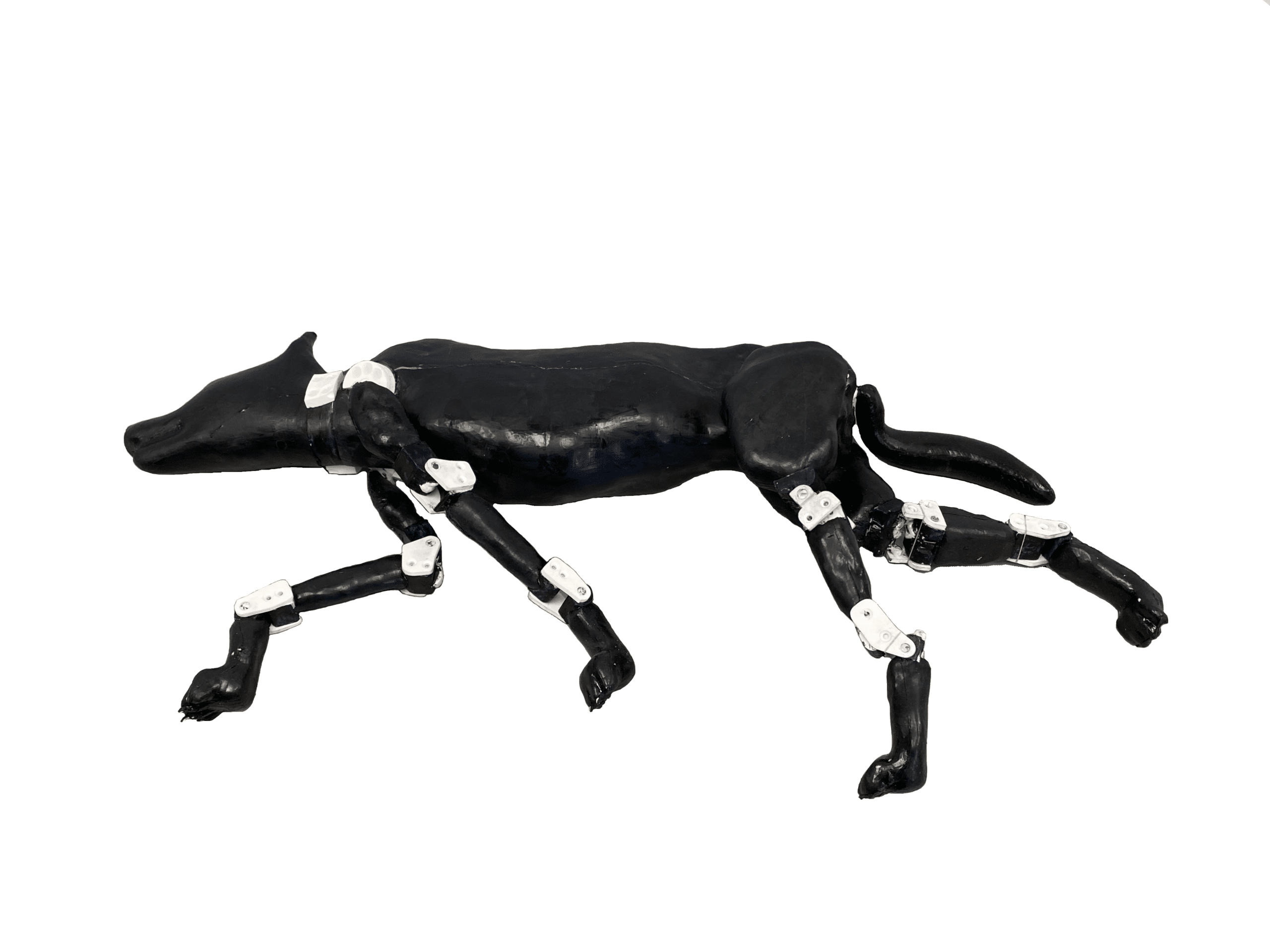
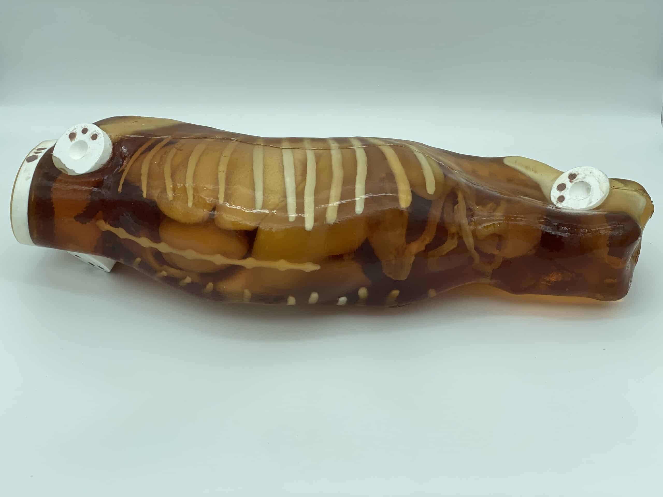
The newly designed detachable dog phantom is a training simulator independent of external hardware/software. The phantom is compatible with Ultrasound and X-RAY/CT imaging. It has all the right features to be an ideal teaching tool for sonographers, radiographers, veterinary residents, and other vet professionals.
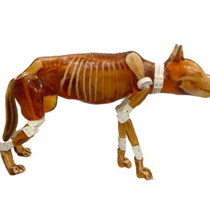
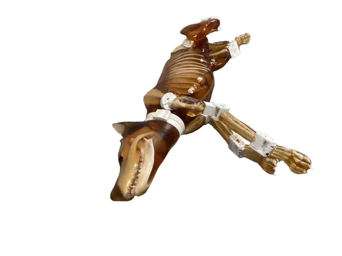
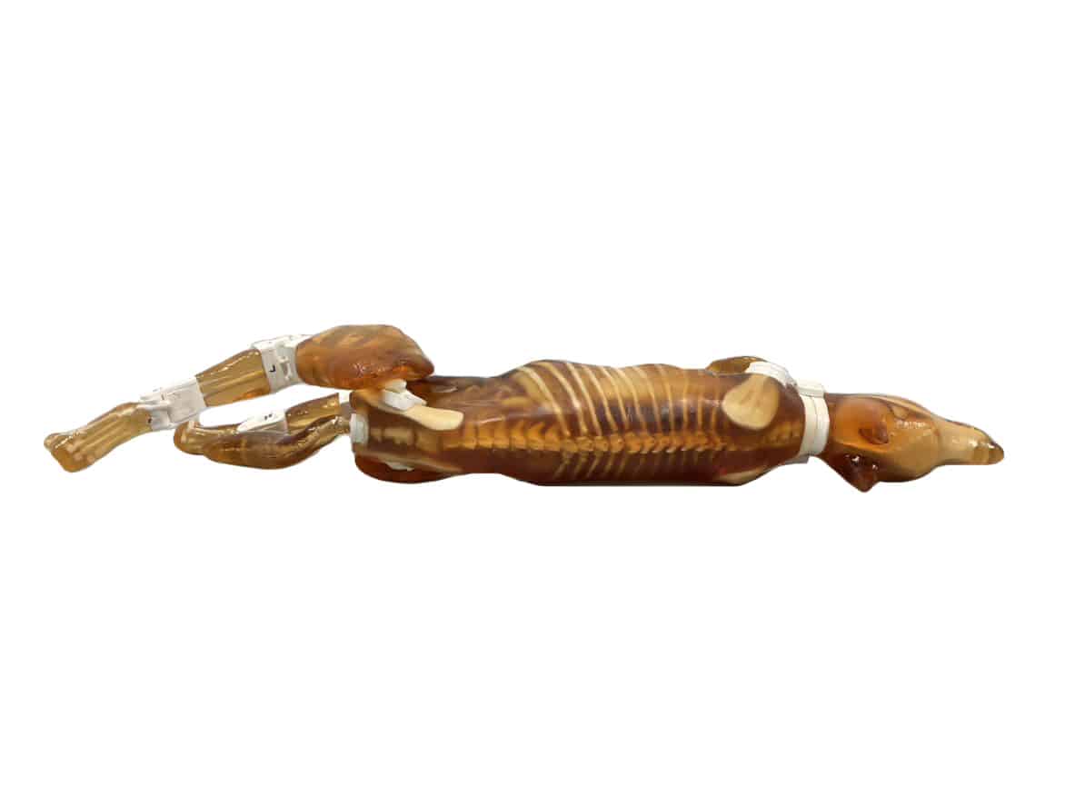
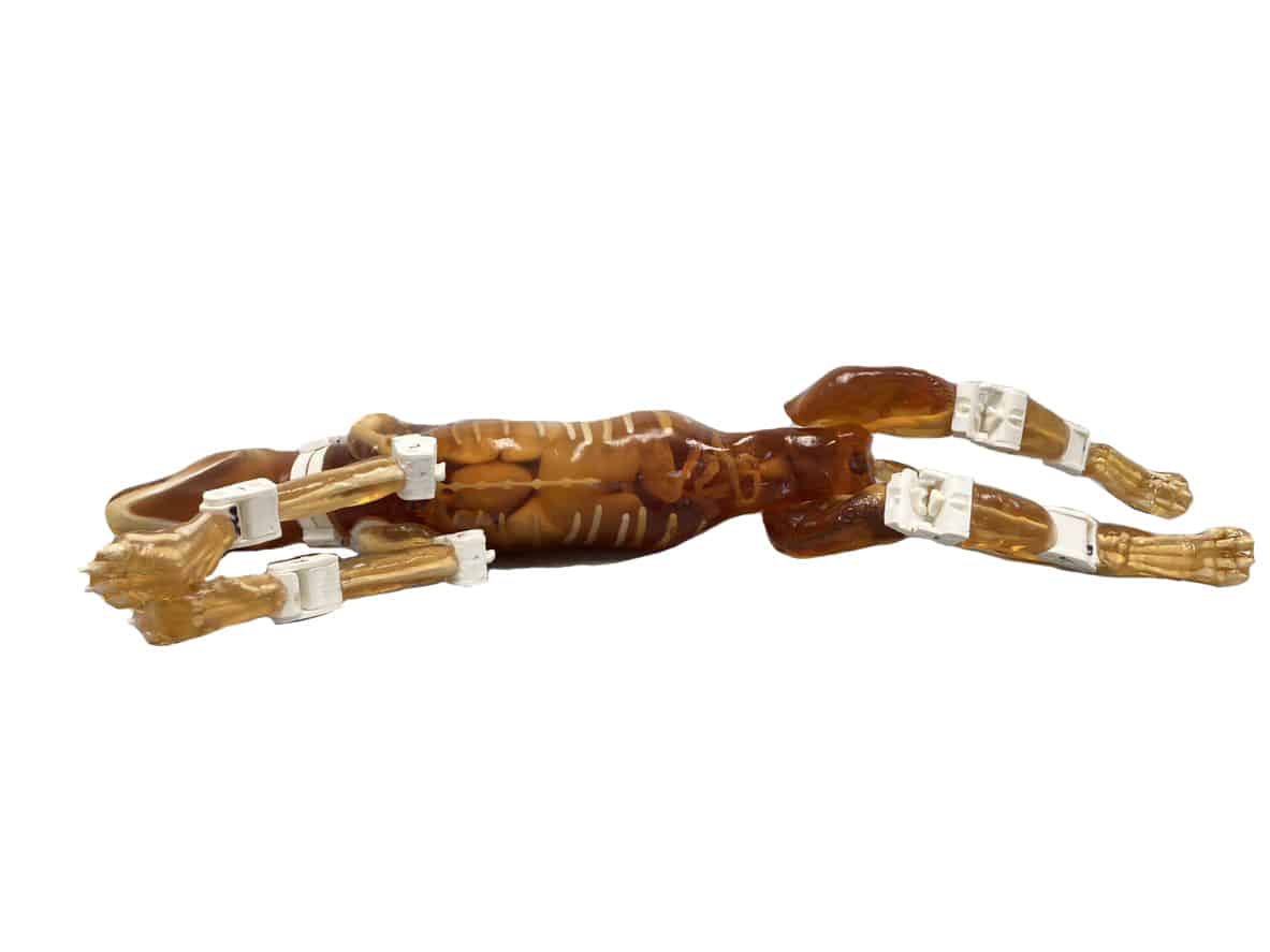
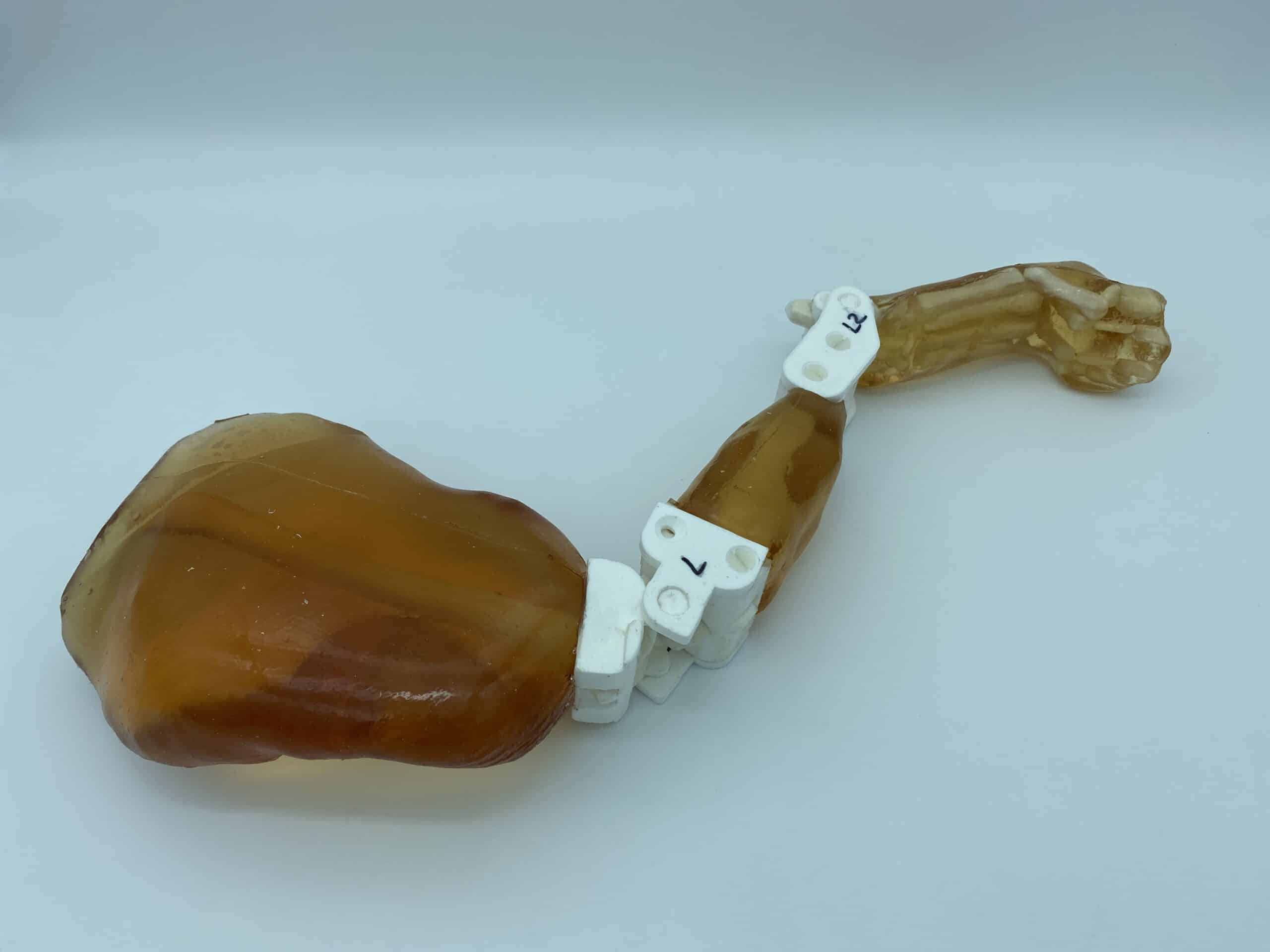
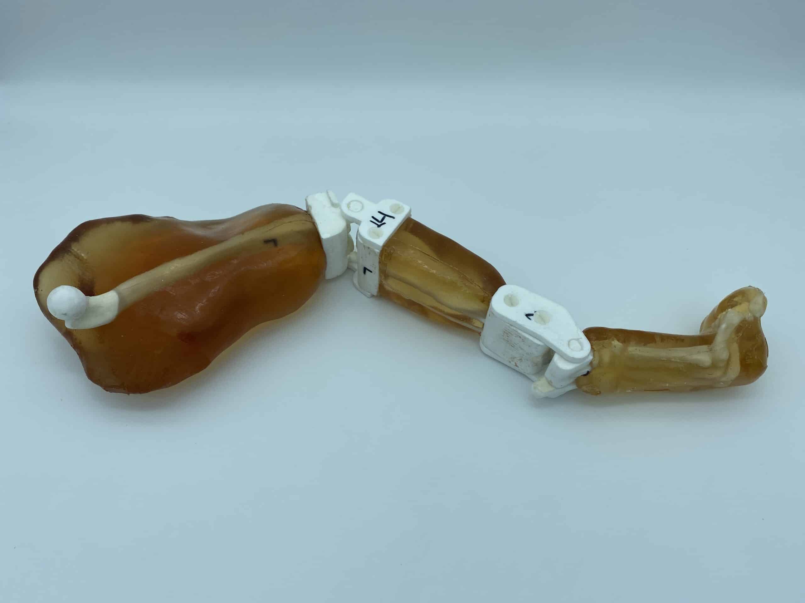
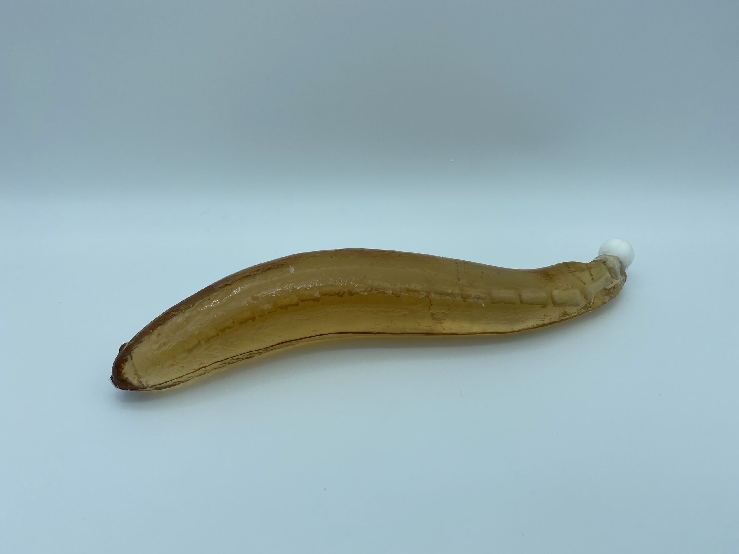
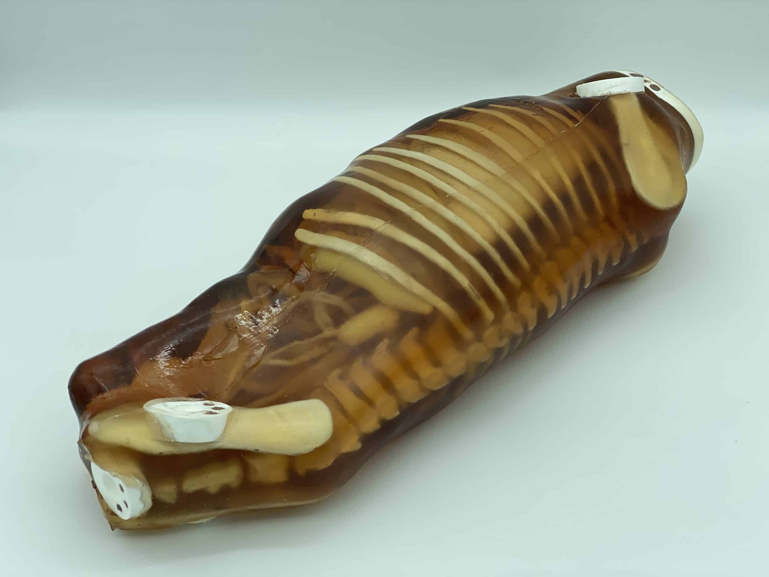
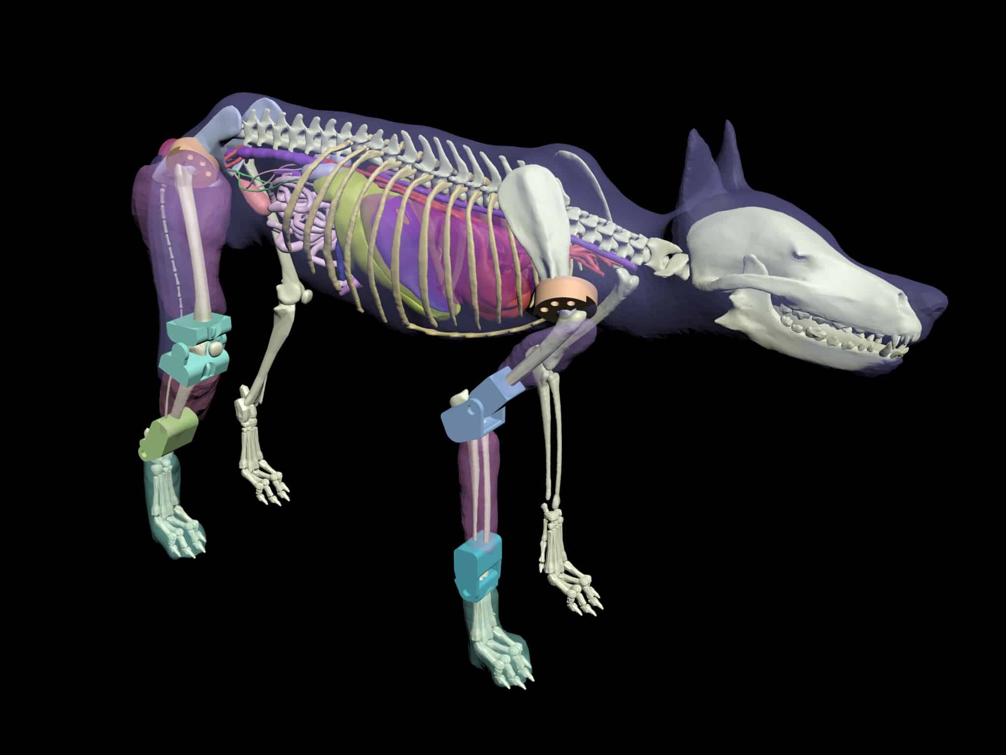
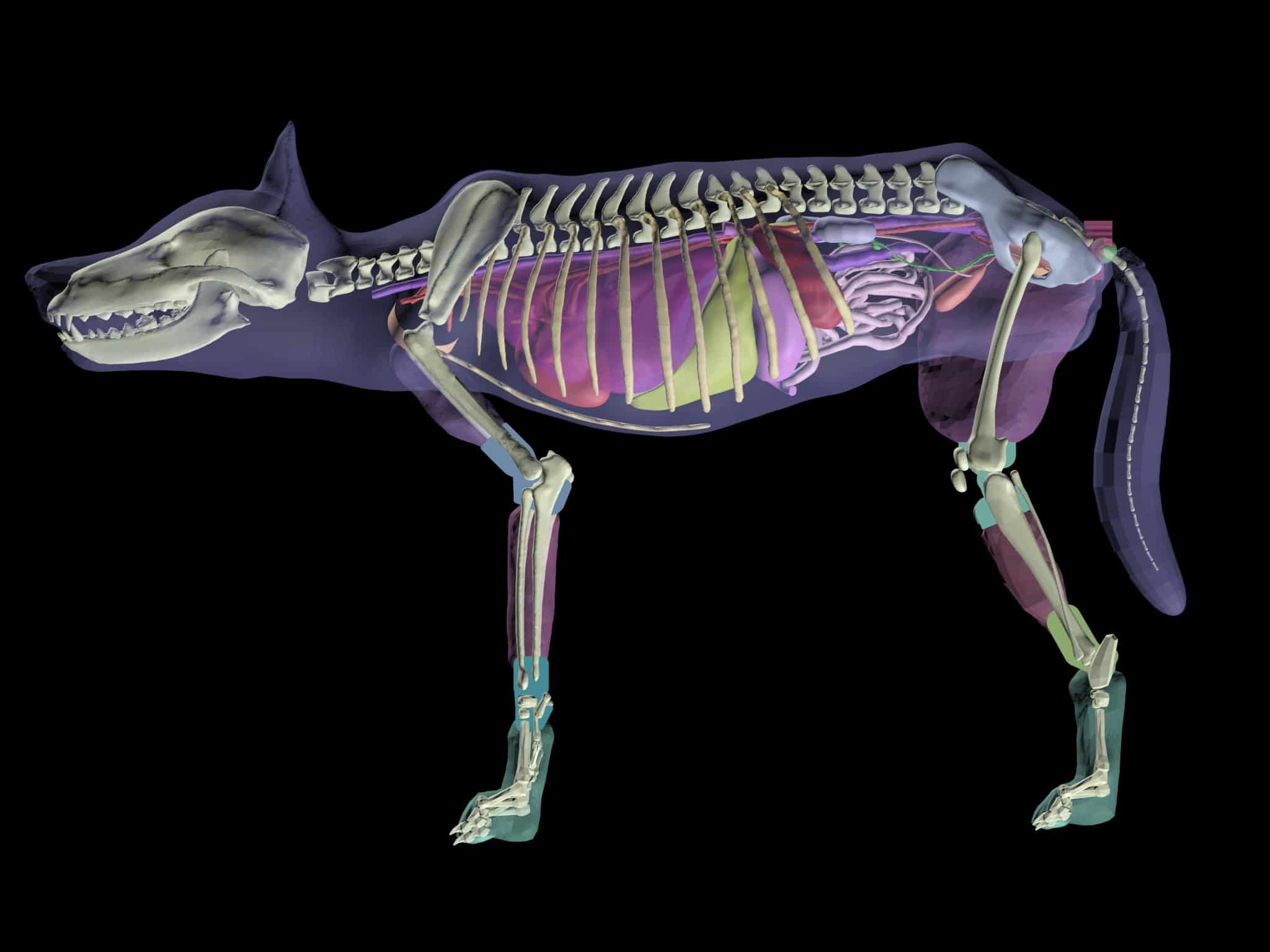
Unlike its predecessor, the new detachable dog phantom has improved anatomical structures and removable body parts (head, limbs, torso, and tail). This improvement is an added advantage as it helps perform various positioning techniques under the US and X-RAY/CT imaging modalities.
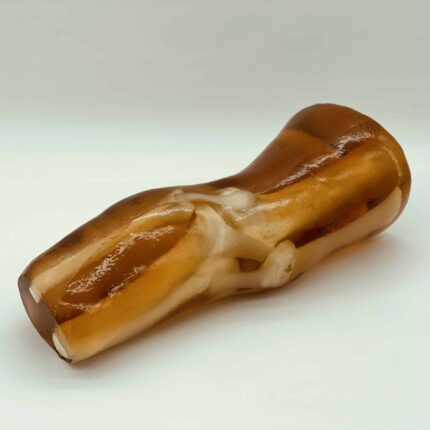
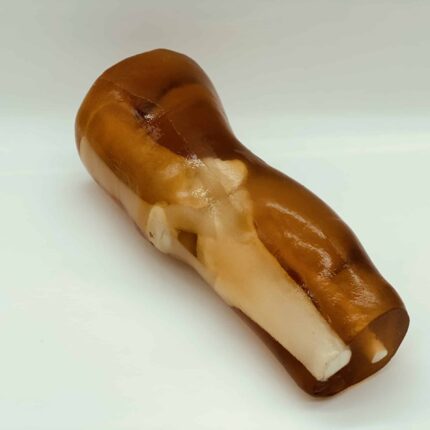
This product is available in two versions: Opaque and Translucent Amber.
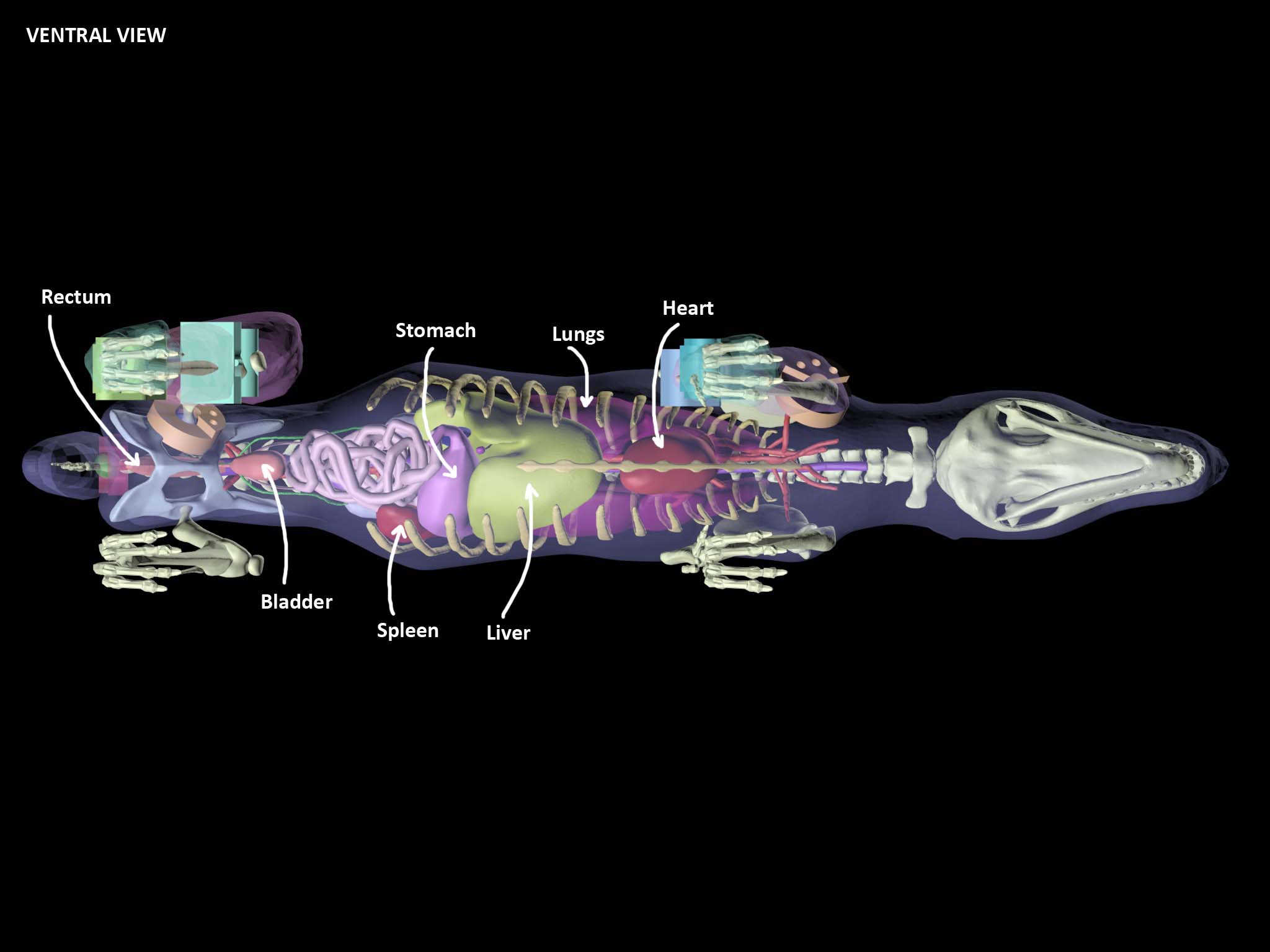
Anatomy
The following anatomical features are included in the standard version of this phantom. Should you require additional organs or components, please reach out to us and we can customize the model to suit your specific needs.
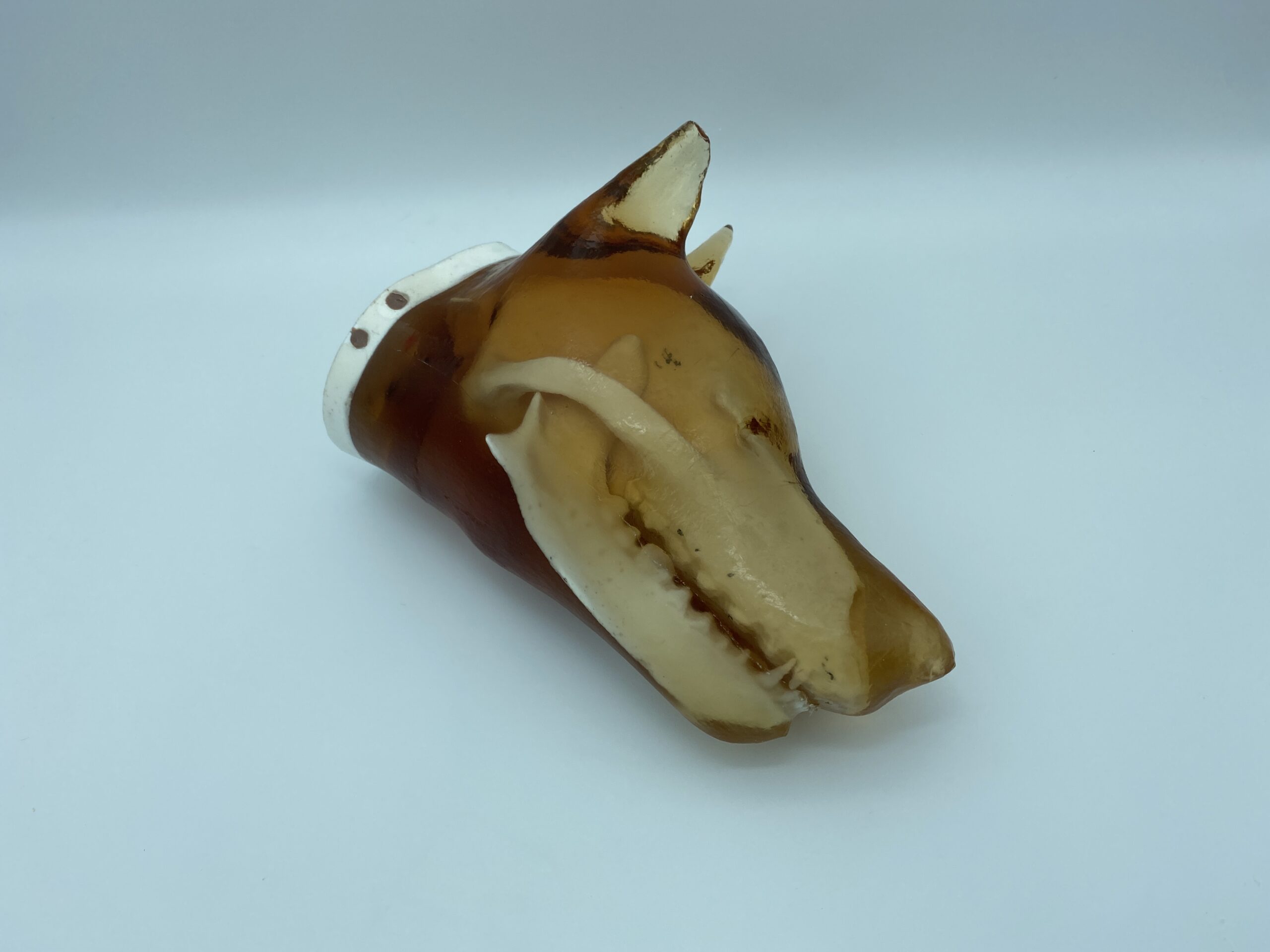
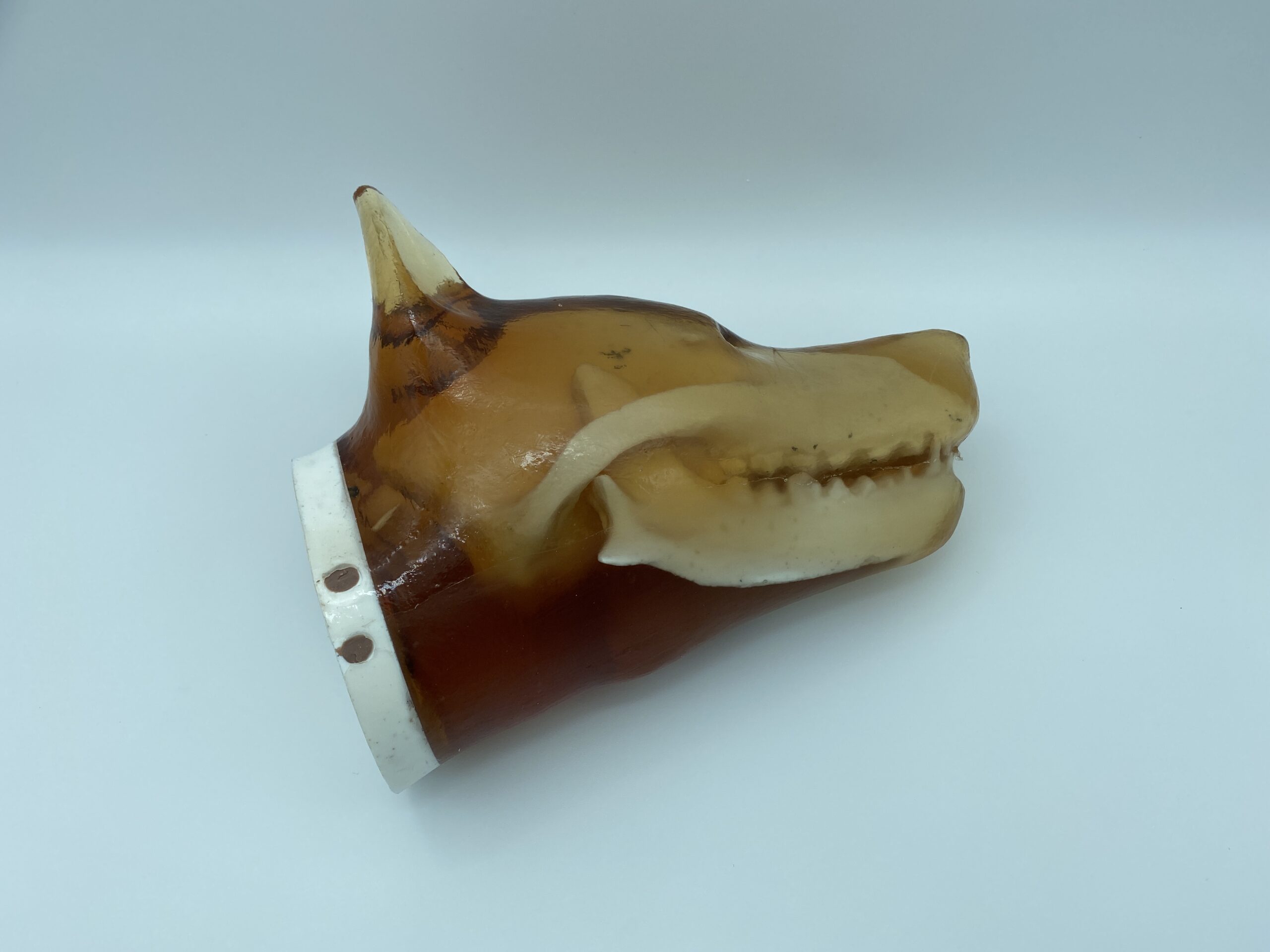
- Adult Dog Head
Dog Skull
Jaw Bone
Realistic Dog Brain
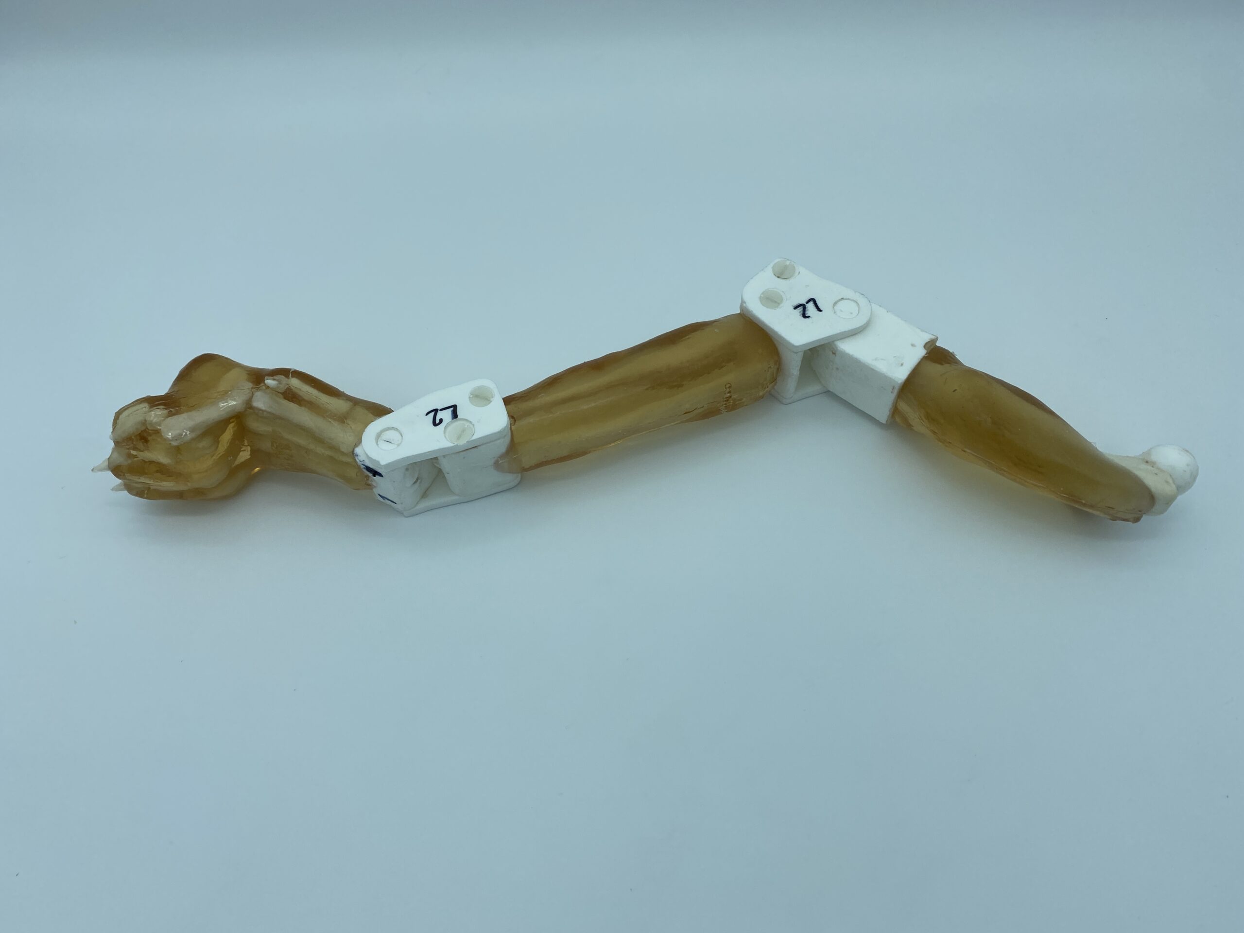
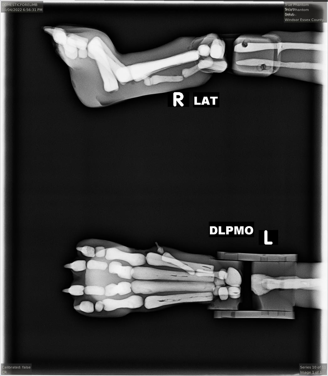
Adult Dog Front Arms
Arm (Humerus)
Elbow Joints
Forearm (Radius, Ulna)
Paw (Wrist with Fingers)
Skin-Mimicking Material
Adult Dog Rear Legs
- Thigh (Femur)
- Knee Joints
- Leg (Tibia, Fibula)
- Paw (Finger Bones)
- Skin-Mimicking Material
Adult Dog Tail
- Tail Bone
- Skin-Mimicking Material
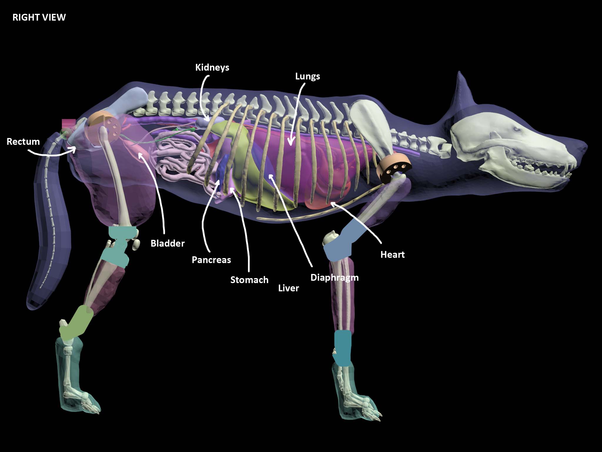
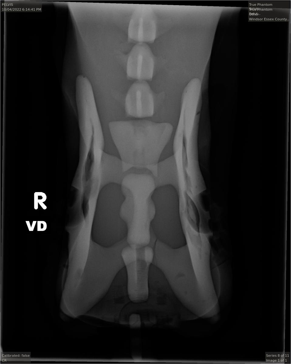
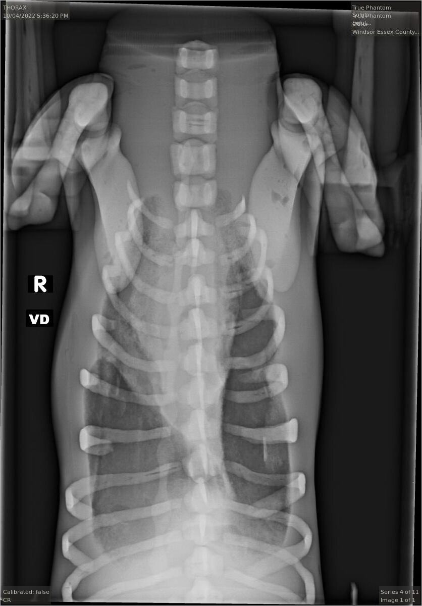
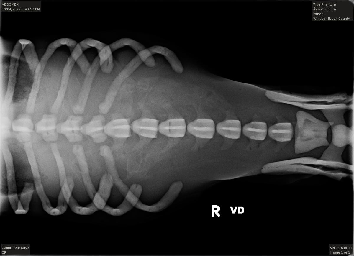
Adult Dog Torso
- Complete Spine
- Complete Ribcage
- Shoulder (Scapula Bones)
- Pelvis
- Heart
- Blood Vessels Connected to Heart
- Lungs
- Diaphragm
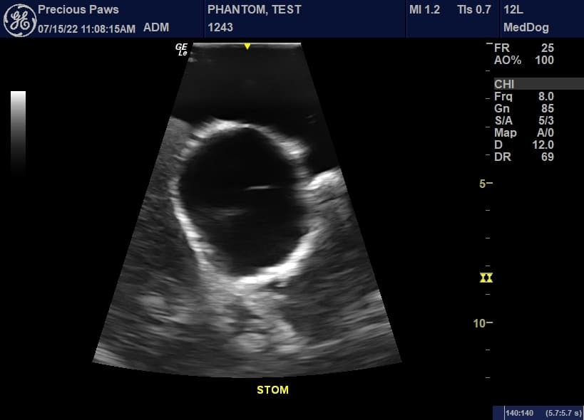
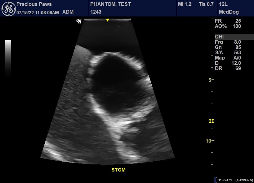
- Stomach
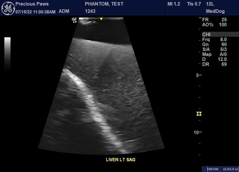
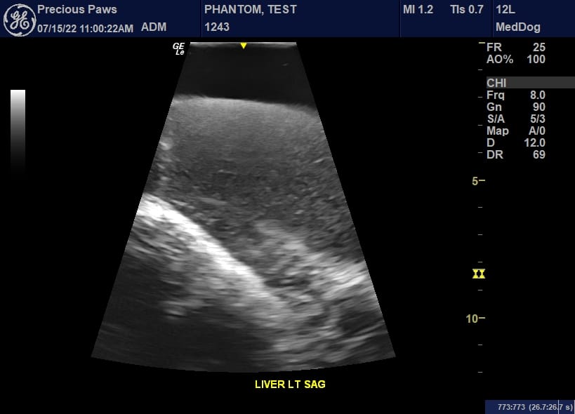
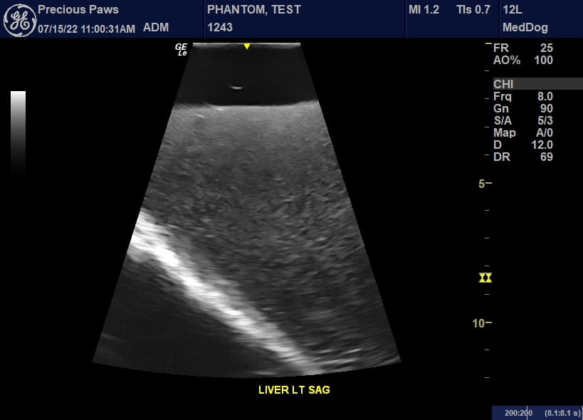
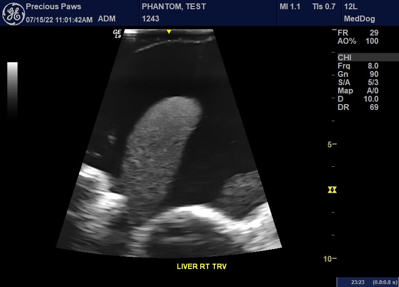
- Liver and Gallbladder
- Kidneys
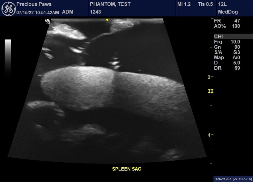
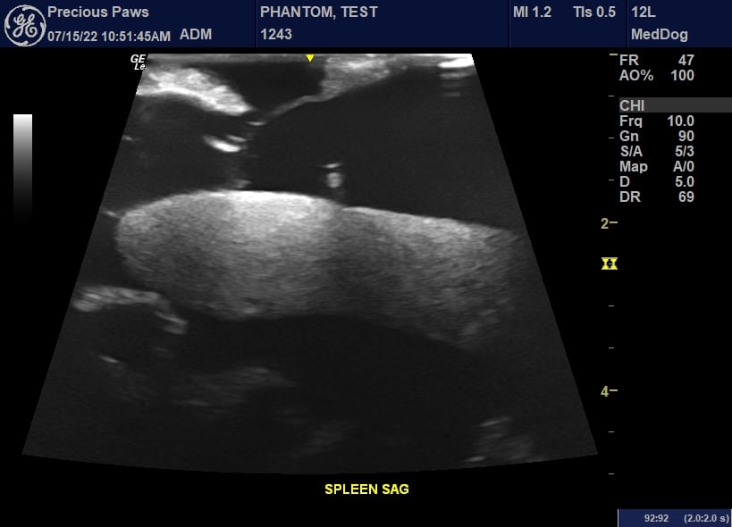
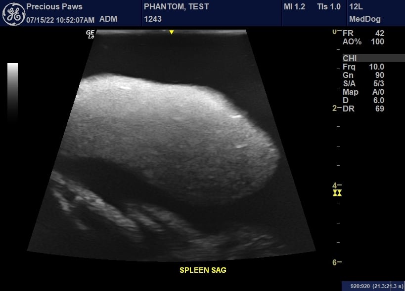
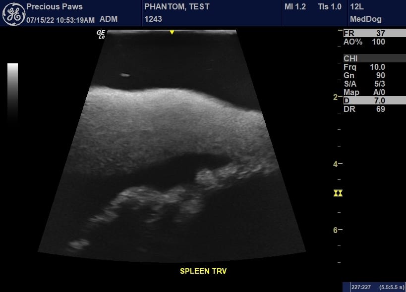
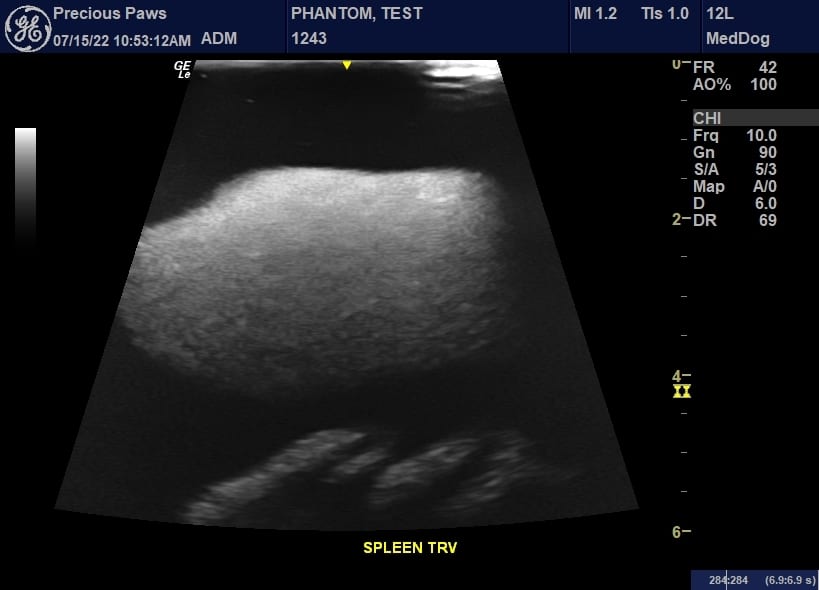
- Spleen
- Pancreas
- Arteries for Kidneys
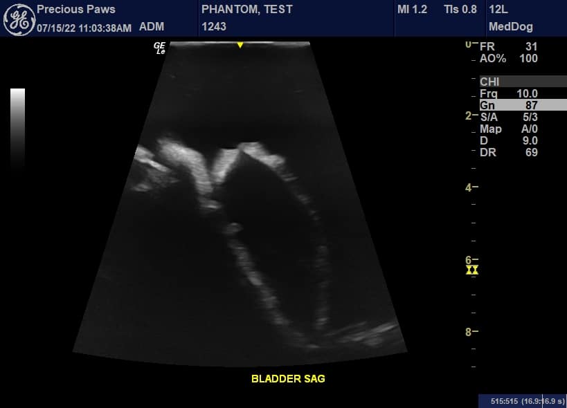
- Bladder
- Ureters and Uterine
- Large and Small Intestine
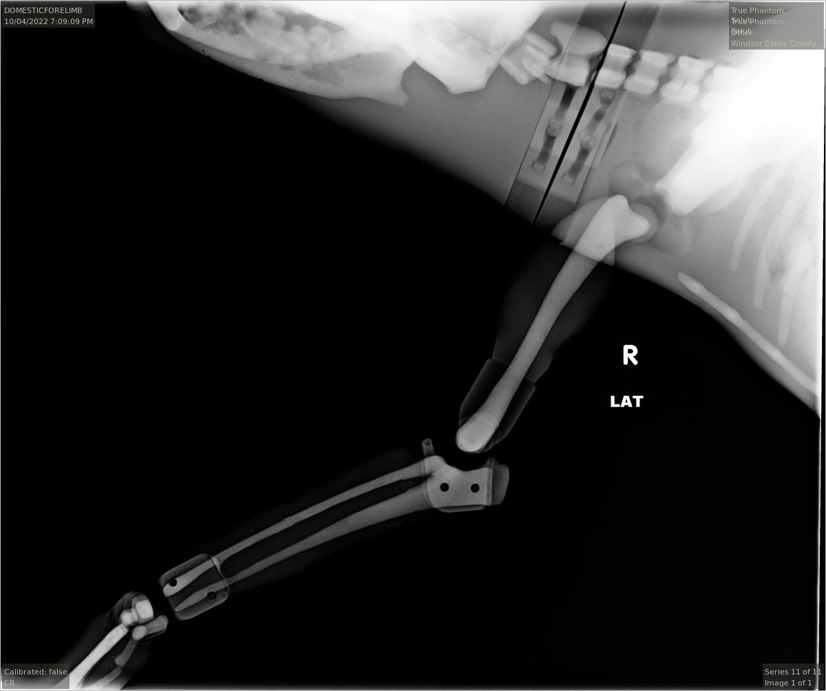
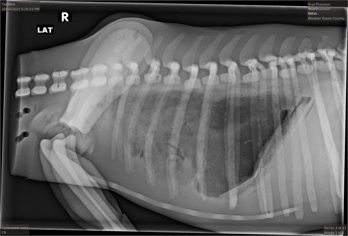
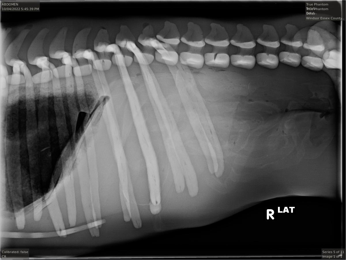
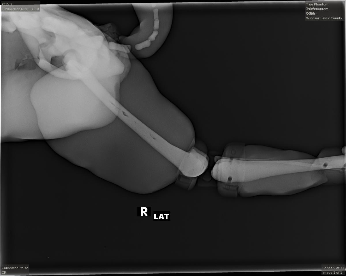
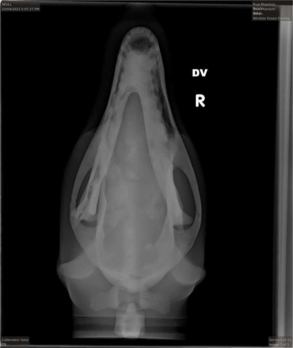
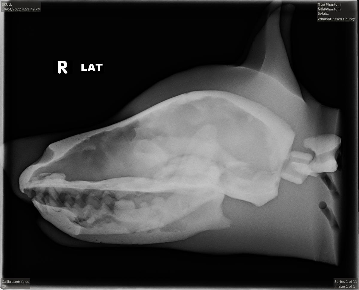
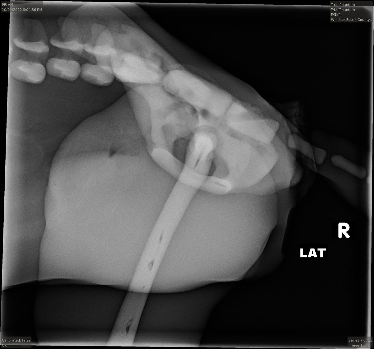
The above X-RAY images were obtained from a standard phantom of our DG-A01 model. An IMAGE VUE CR 20 X-RAY Scanner was used to scan the phantom. This phantom was scanned and validated by Elyse Morassutti, a registered Veterinary technician from the Windsor-Essex County Humane Society
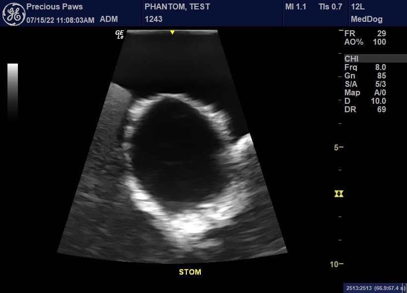
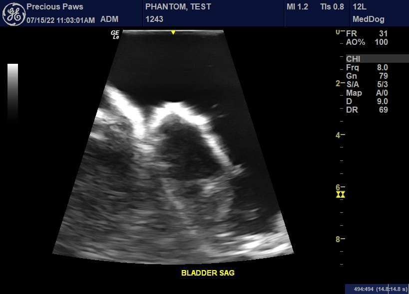
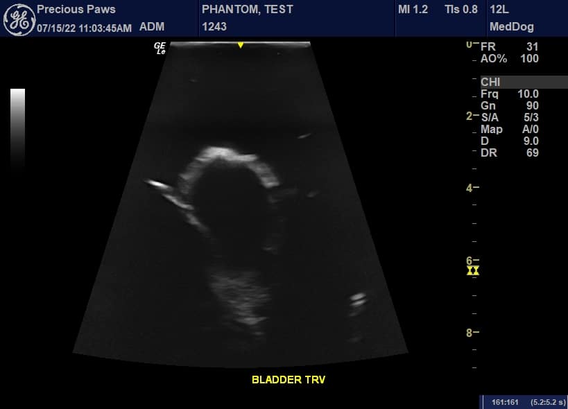
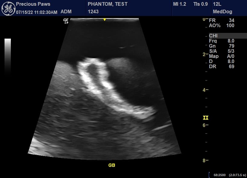
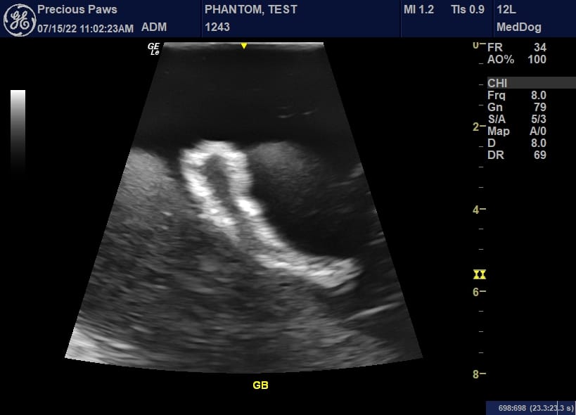
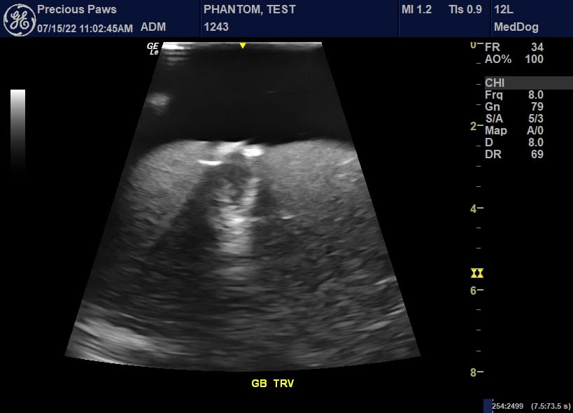
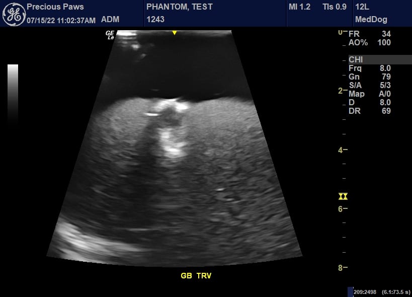
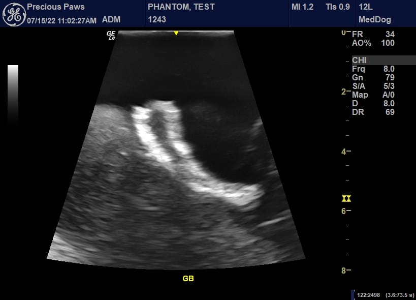
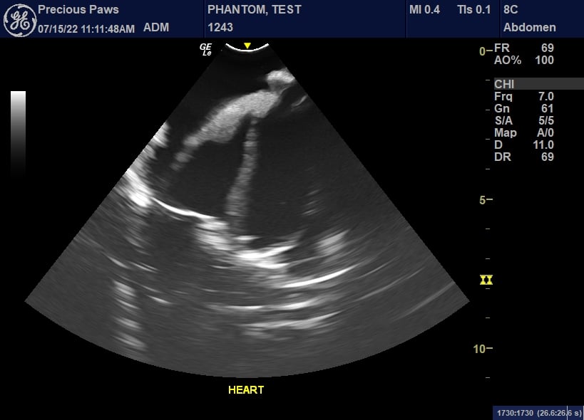
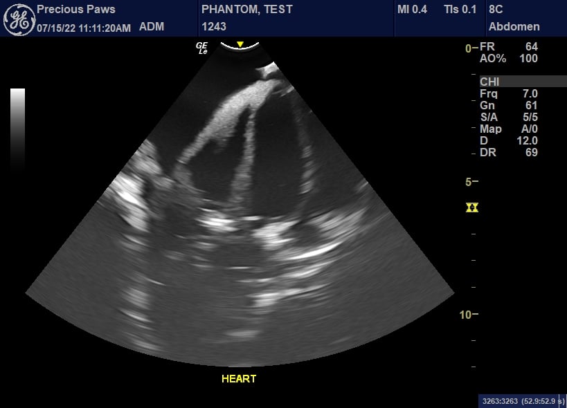
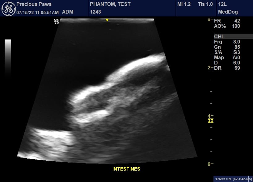
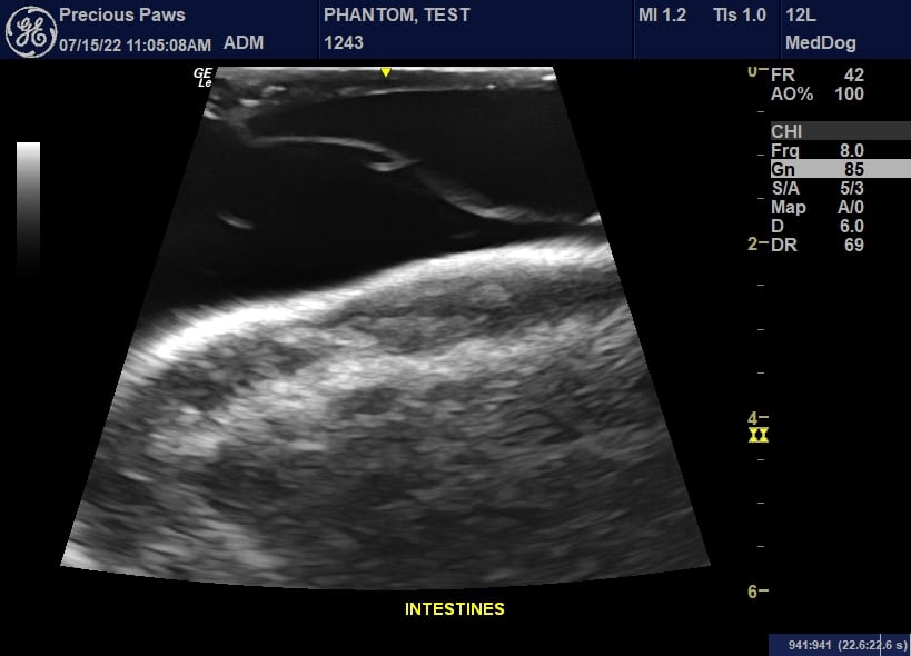
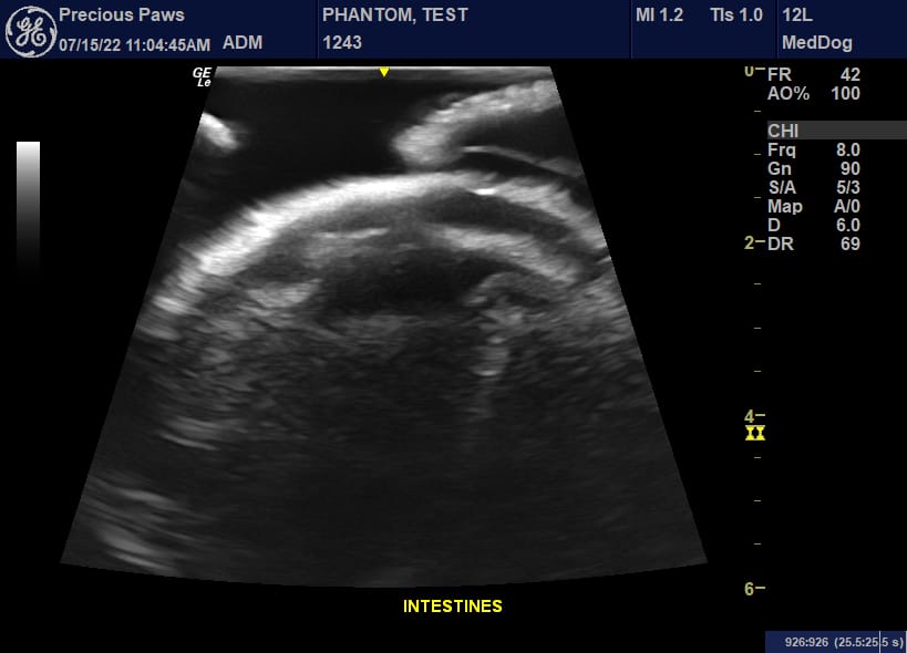
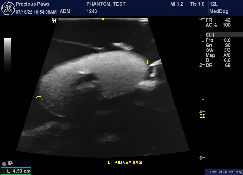
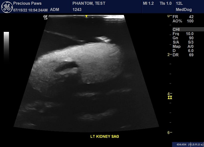
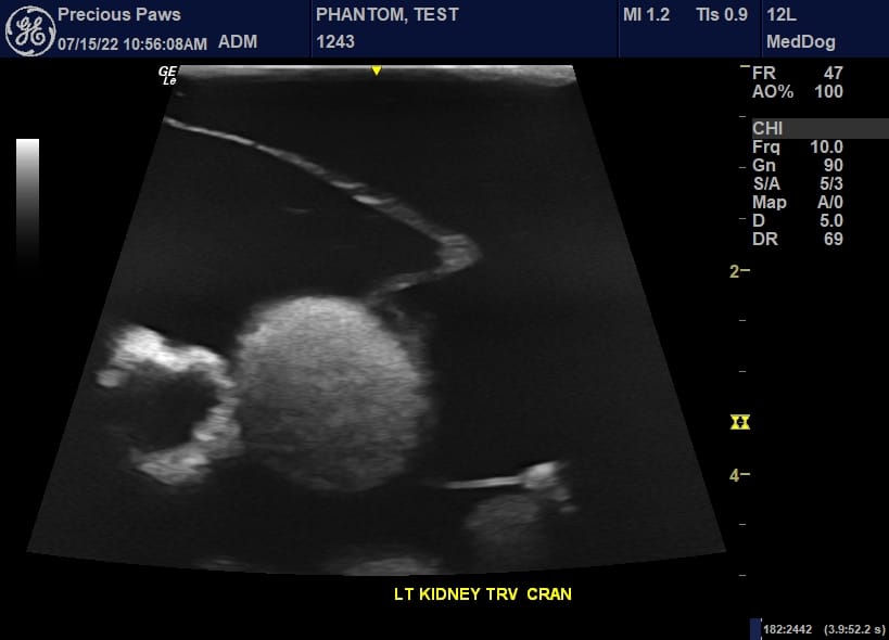
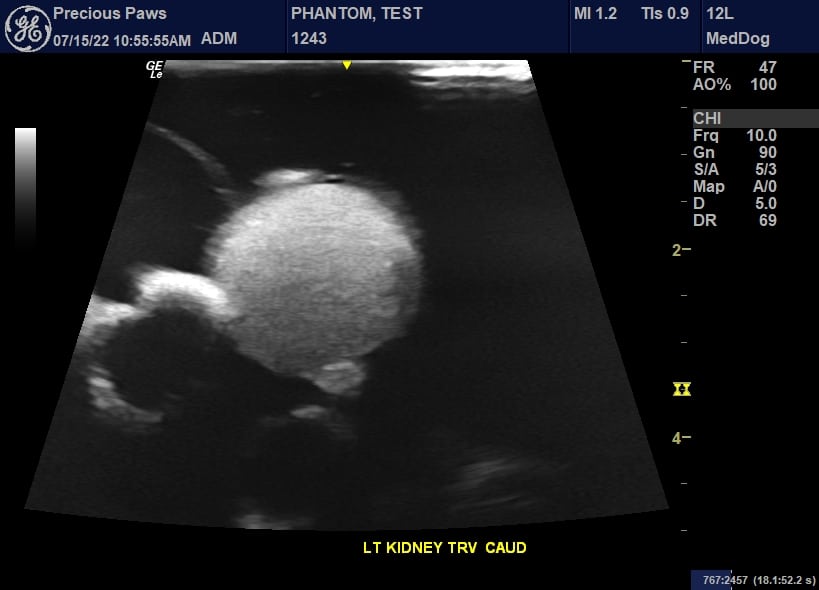
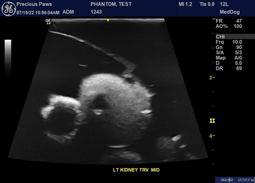
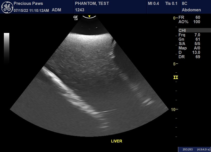
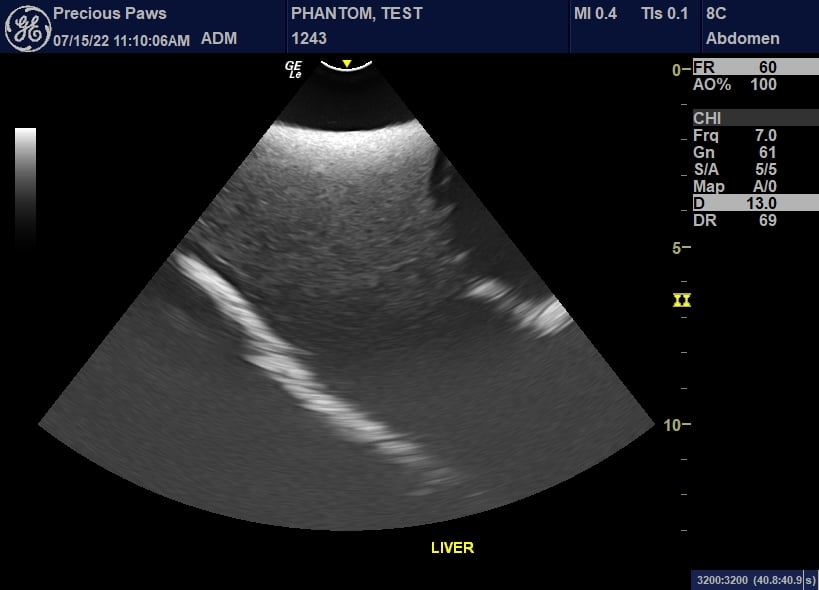
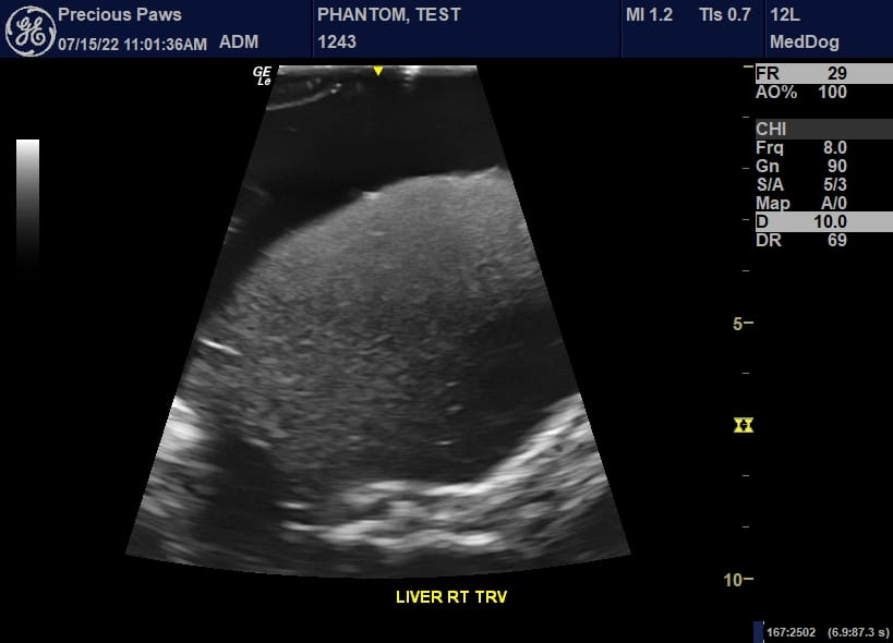
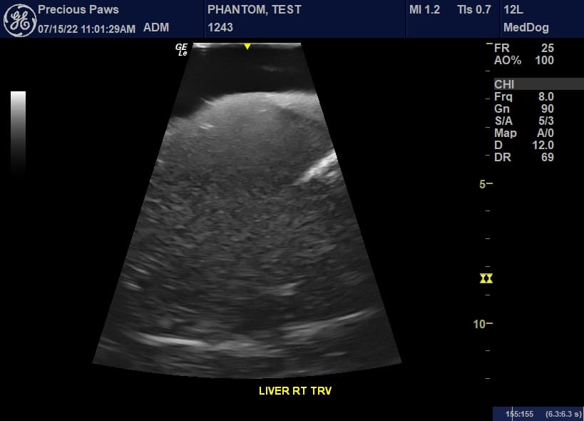
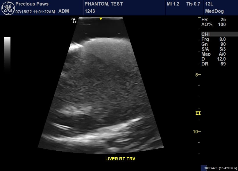
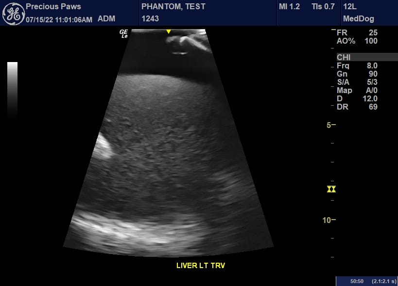
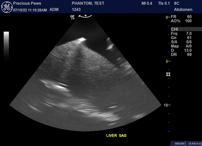
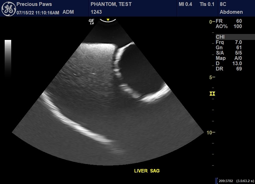
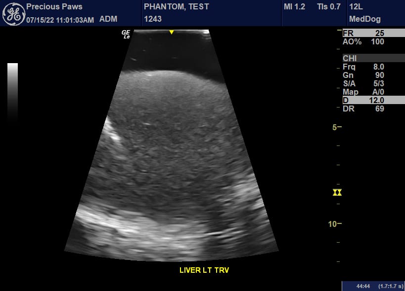
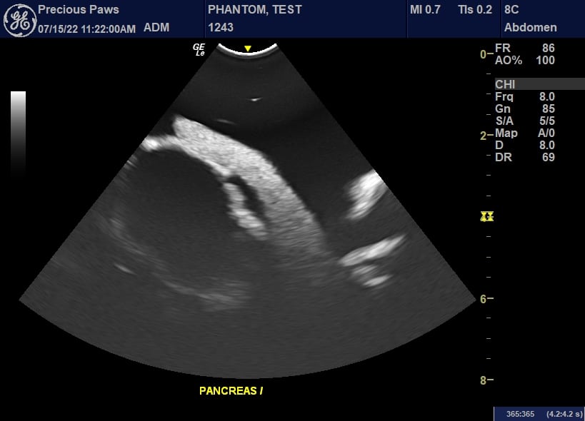
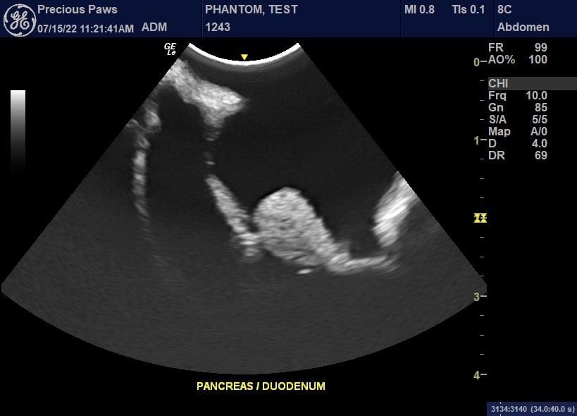
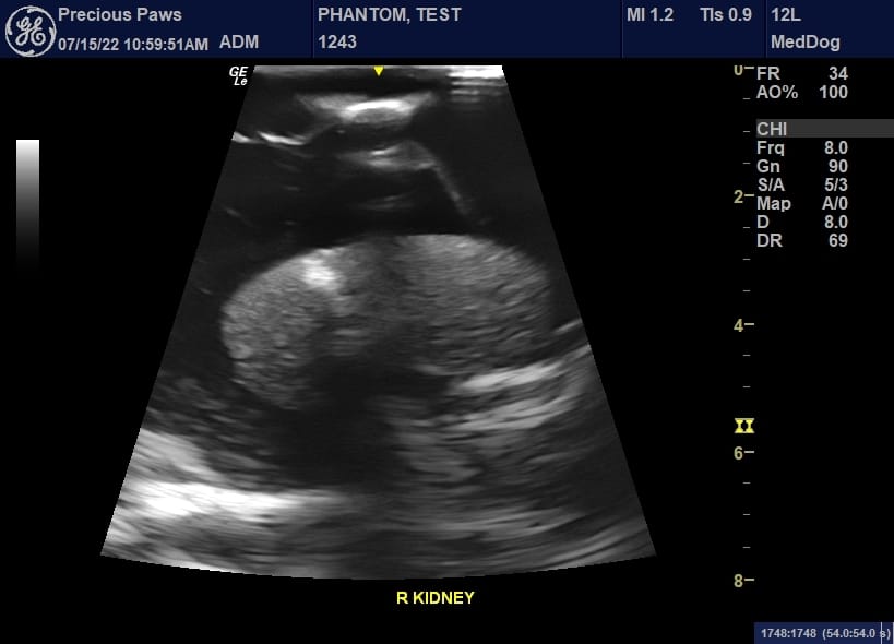
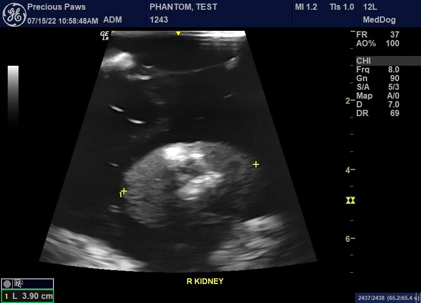
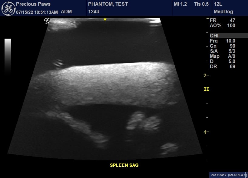
*/The above results were obtained from a transparent model of DG-A01. All ultrasounds were performed with the GE Logiq e R8 using the standard 8C micro convex and the L12 Linear probes. This allowed us to interrogate and obtain the best-quality images. By having two different ultrasound probes with varying frequencies, we were able to switch our probes throughout the exam while assessing different organs/body parts resulting in obtaining high-quality images. The phantom was scanned and validated by a VET ultrasound specialist from Precious Paws Ultrasound./*
Technical Properties:
| Type of Tissue | Sound Velocity[m/s] | Density [g/cm3] | Attenuation Measured at 2.25 MHz [dB/cm] | Hardness [Shore OO] | T2 [ms] | Speckles |
| Organs with Speckles (liver, kidneys, etc.) | 1400 ± 10 | 0.99 | 1.0 ± 0.2 | 20 | 70 | VARIABLE |
| Organs without Speckles (stomach, intestines, etc.) | 1400 ± 10 | 0.99 | 1.0 ± 0.2 | 20 | 70 | NO |
| Body Tissue | 1400 ± 10 | 1.00 | 1.2 ± 0.2 | 30 | 65 | NO/LOW |
| Cortical Bone | 3000 ± 30 | 2.31 | 6.4 ± 0.3 | N/A | N/A | N/A |
| Trabecular Bone | 2800 ± 50 | 2.03 | 21 ± 2 | N/A | N/A | N/A |
| Brain Matter | 1400 ± 10 | 0.99 | 1.0 ± 0.2 | 20 | 70 | YES |
| Thermal Properties of our Bone-Mimicking Material | |||||
| Thermal Conductivity | Volumetric Specific Heat Capacity | Thermal Diffusivity | Thermal Resistivity | Specific Heat | Speed of Sound |
| 0.776 W/ m K | 1.040 MJ/ m^3 K | 0.746 mm^2/ s | 1.289 m K/ W | 0.978 J/ g Deg Celsius | |
| HU Values of the Tissue Mimicking-Materials | ||
| S.No. | Tissue Type | HU Value (Average) |
| 1 | Body Tissue | -25 |
| 2 | Brain Tissue | -25 |
| 3 | Trabecular Bone | 800 |
| 4 | Cortical Bone | 1300 |
| 5 | Aorta | 40 |
| 6 | Vena Cava | 40 |
| 7 | Trachea | 80 (Tissue Part), -1000 (Air Filled Part) |
| 8 | Pancreas | 110 |
| 9 | Spleen | 110 |
| 10 | Kidneys | 110 |
| 11 | Bladder | 35 |
| 12 | Rectum Wall | 100 |
| 13 | Sigmoid Colon Wall | 100 |
| 14 | Heart | 40 |
| 15 | Liver | 110 |
| 16 | Gallbladder | 35 |
View our technical properties, thermal properties of bone-mimicking tissue, and HU values of the tissue-mimicking material in the tables accessible through the button below.
Dog Phantom
User Manual/Assembly Instructions
2-Year Warranty & Unlimited Customer Service
Hard Carry Case
Phantom:
- Size: 33 x 18 x 6 Inches
- Weight: 40 Lbs (approx.).
Shipment:
- Size with Hard Carry Case: 32 x 23 x 13 Inches
- Weight: 99 Lbs (approx.)
- Soft tissue and organs: Composition of urethane-based soft resin
- Synthetic bones: Patented ceramic-reinforced epoxy-based composite material